

今週、JSAP Expo Autum 2024 で NanoAndMore Japan にお越しくださいMon Sep 16 2024
NanoAndMoreジャパンは応用物理学会 秋季学術講演会 併設展示会 JSAP EXPO Autumn - this is the main text
2024に出展しています。
主催:公益社団法人 日本応用物理学会
日時:2024年9月16日 (月)-9月20日(金)
場所:新潟市 朱鷺メッセ ブーズ番号I-24
https://meeting.jsap.or.jp/
#AFMカンチレバー #AFMプローブ #日本応用物理学会 #応用物理学 #物理学 #SPMプローブ #原子間力顕微鏡 #走査型プローブ顕微鏡 #jsap #超高周波AFMプローブ #マイクロカンチレバー #ナノテクノロジー #材料科学 #生物物理学 #応用物理学 #走査型プローブ顕微鏡 学 #応用物理学 #走査型プローブ顕微鏡

Deformable microlaser force sensingThu Sep 12 2024
#Mechanicalforces are key regulators of #cellularbehavior and function, affecting many fundamental #biologicalprocesses such as #cellmigration, embryogenesis, immunological responses, and pathological states.*
Specialized #forcesensors and imaging techniques have been developed to quantify these otherwise invisible forces in #singlecells and #invivo. However, current techniques rely heavily on high-resolution microscopy and do not allow interrogation of optically dense tissue, reducing their application to 2D cell cultures and highly transparent #biologicaltissue.*
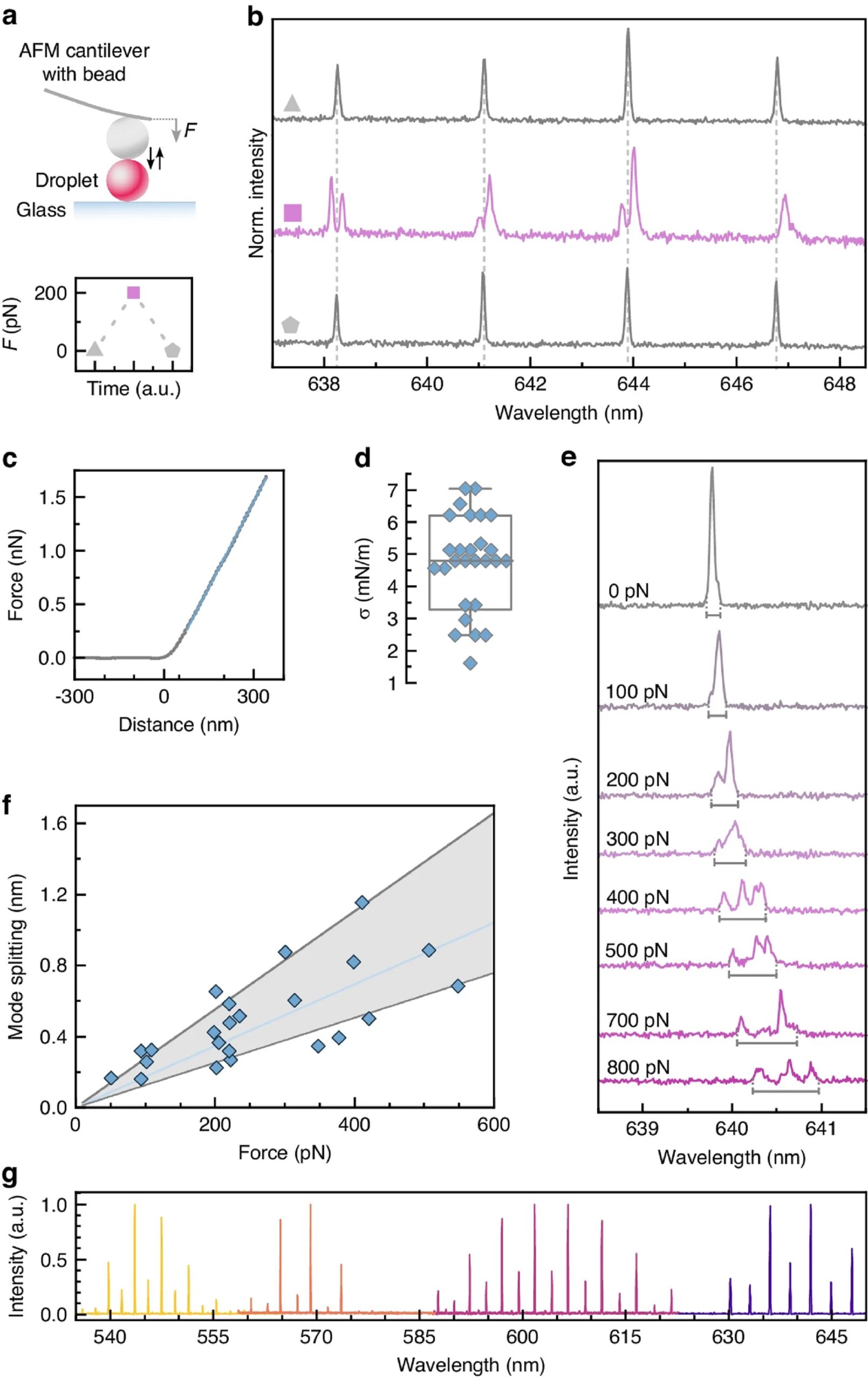
In the article “Deformable microlaser force sensing” Eleni Dalaka, Joseph S. Hill, Jonathan H. H. Booth, Anna Popczyk, Stefan R. Pulver, Malte C. Gather and Marcel Schubert introduce DEFORM, deformable microlaser force sensing, a spectroscopic technique that detects sub-nanonewton forces with unprecedented spatio-temporal resolution.
DEFORM is based on the spectral analysis of laser emission from dye-doped oil microdroplets and uses the force-induced lifting of laser mode degeneracy in these droplets to detect nanometer deformations.
The authors use gold-standard #atomicforcemicroscopy ( #AFM) measurements to validate the absolute scale of the extracted forces and to evaluate the optical response of individual droplets in order to be able to demonstrate the ability of DEFORM to extract absolute mechanical forces in a later step.
The controlled deformations of single droplet microlasers were performed using an #atomicforcemicroscope which was installed on the inverted microscope that was used for the lasing experiments, allowing simultaneous optical and mechanical characterization of the droplets.
For #indentation, a 17 µm glass sphere was glued to the #AFMtip of a soft #AFMcantilever with nominal stiffness of k = 0.01 N/m ( NANOSENSORS™ uniqprobe qp-SCONT). https://www.nanosensors.com/uniqprobe-uniform-quality-soft…
Eleni Dalaka et al. also developed a model that links changes in laser spectrum to applied force and allows to extract the eccentricity of the flexible microlaser droplets. Based on the known deformation of the microlasers, the applied force can then be directly calculated.
NANOSENSORS™ uniqprobe #AFMprobes offer an outstanding uniformity of the mechanical AFM cantilever characteristics which is particularly important for applications where a large number of AFM probes with known and near identical force constants or resonance frequencies are needed. The AFM cantilevers of the uniqprobe series are especially adapted for applications in #molecularbiology, #biophysics and #quantitativenanomechanicalstudies .
Please have a look at the NANOSENSORS blog for the full citation and a direct link to the full article

An explicit model to extract viscoelastic properties of cells from AFM force-indentation curvesFri Sep 06 2024
The #viscoelasticbehavior of #softmaterials, especially #cells and #tissues, has been extensively investigated due to its importance in many biological and physiological processes that take place during development and even disease.*
The #atomicforcemicroscope ( #AFM), in particular, is still nowadays one of the most popular methods to quantify the #mechanicalproperties of cells, due to its conformity with various material types and geometries and the rather simple analysis process of the material properties.*
For a typical #AFMindentationmeasurement, an #AFMcantilever, with a distinct #AFMtip shape, moves toward the sample with a predefined velocity and indents it until a prescribed force is reached. Generally, a Hertzian model is fitted to the approach part of the force-indentation curves to quantify the apparent Young's modulus.*
Cells and tissues, however, show not only elastic but also #viscousbehavior that is evident from the hysteresis between the approach and retraction segments of the #forceindentationcurve. Consequently, assessing this viscoelastic behavior is imperative for understanding the complex nature of #biologicalmatter.*
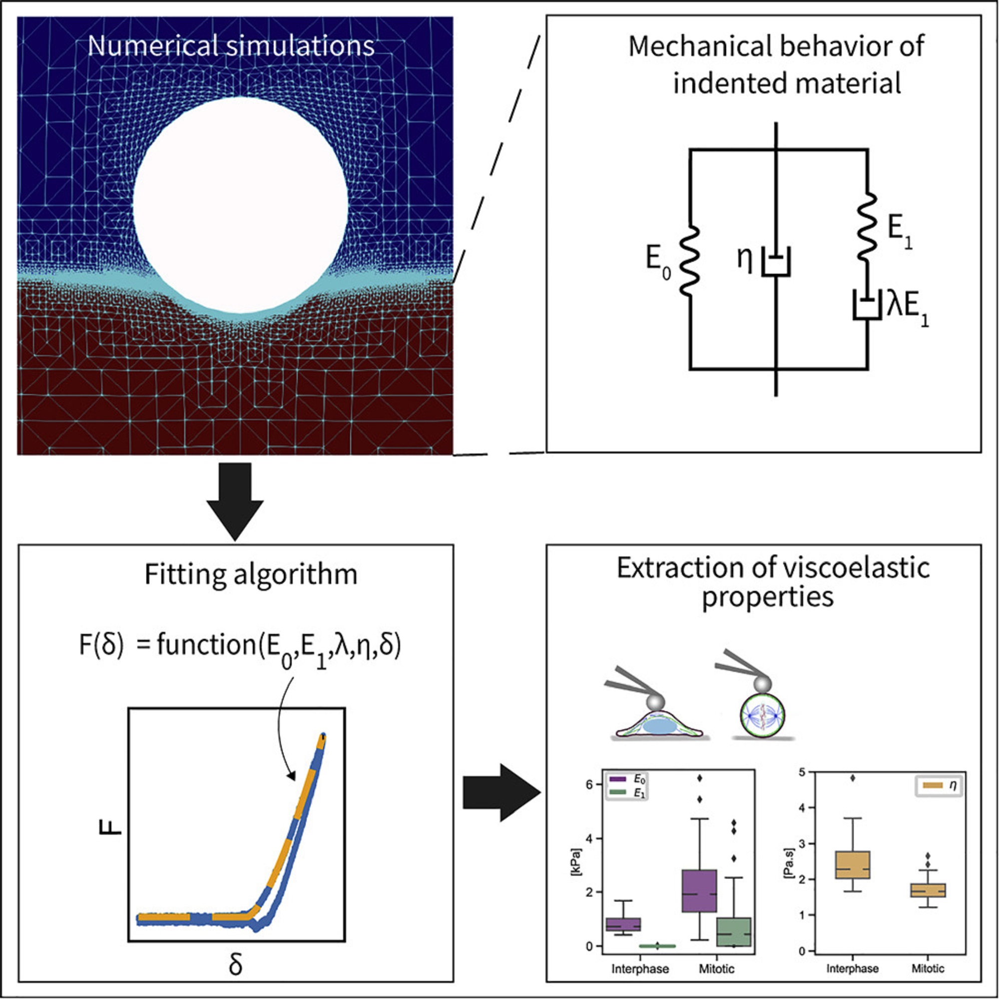
In the article “An explicit model to extract viscoelastic properties of cells from AFM force-indentation curves”, Shada Abuhattum, Dominic Mokbel, Paul Müller, Despina Soteriou, Jochen Guck and Sebastian Aland propose a new fitting model to extract the viscoelastic properties of soft materials from #AFMforceindentationcurves. *
Shada Abuhattum et al. show that the proposed Kelvin-Voigt-Maxwell (KVM) model adequately captures the force-indentation curves of materials having different mechanical characteristics. *
Based on the simulation results, Shada Abuhattum et al. further propose an explicit force-indentation relation to be fitted to the #forceindentationcurves. This explicit relation simplifies the association of the mechanical properties with physically meaningful components and processes.
Finally, the authors apply the fitting model to a number of samples, including poroelastic and viscoelastic #hydrogels as well as #HeLacells in two different cell cycle phases, interphase and mitotic. *
Their findings demonstrate that the proposed model can reliably extract viscoelastic properties from conventional force-indentation curves. Moreover, the model is able to assess the contribution of the different elastic and viscous elements, and thus allows a direct comparison between the viscoelastic nature of different materials.*
#AFMmeasurements were preformed using a commercially available Atomic Force Microscope. To indent the samples, NanoWorld Pyrex-Nitride tipless AFM cantilevers PNP-TR-TL https://www.nanoworld.com/pyrex-nitride-triangular-silicon… were modified by gluing 5 μm diameter polystyrene beads to the underside of the AFM cantilevers using two component glue.*
Please have a look at the NanoWorld blog for the full citation and a direct link to the full article


We are often asked whether our platinum coated ElectriCont-G AFM probes are suitable for Conductive AFM.Wed Sep 04 2024
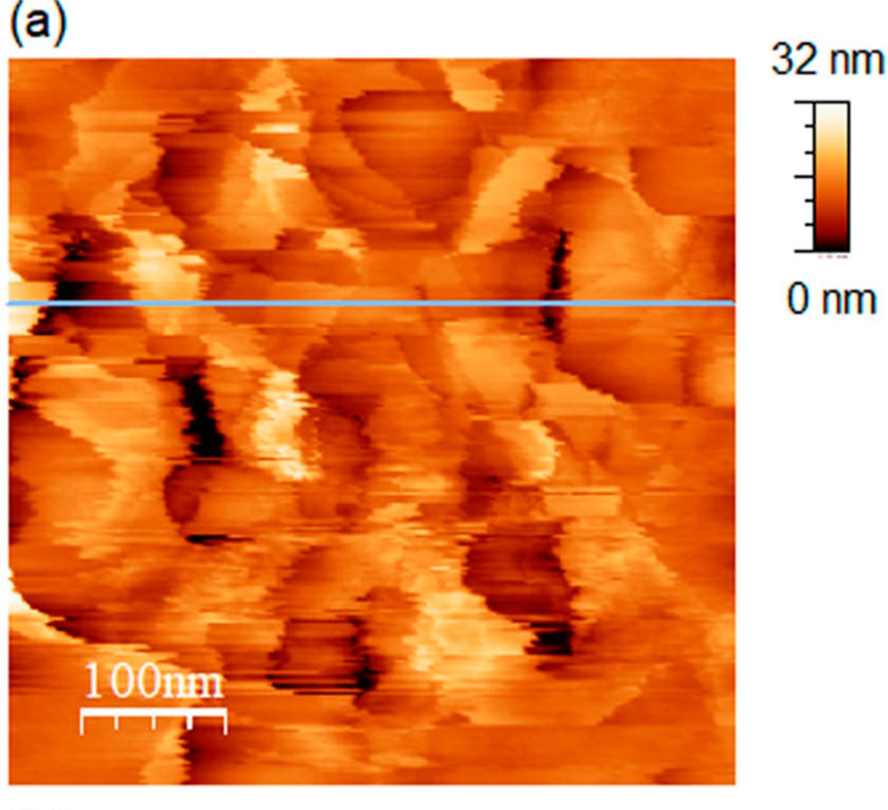
The ElectriCont-G are not designed or recommended for C-AFM, although some of our customers use them as a cost-effective alternative to conductive diamond coated AFM probes. Fine-tuning of scan and electrical parameters is critical to preserve the platinum coating in this demanding AFM application.
The image shows two C-AFM scans on a partially processed semiconductor device. Throughout the left scan the C-AFM current remains stable. In the right scan the current gradually drops as the scan progresses indicating fast wear of the platinum coating due to abrasion and/or local heating.
AFM measurements are courtesy of the Institute for Semiconductor Technology and Nanoelectronics, Darmstadt University of Technology.
Light-Induced Ferroelectric Modulation of p-n Homojunctions in Monolayer MoS2Tue Sep 03 2024
The association of #2Dmaterials and #ferroelectrics offers a promising approach to tune the optoelectronic properties of atomically thin #TransitionMetalDichalcogenides (TMDs). *
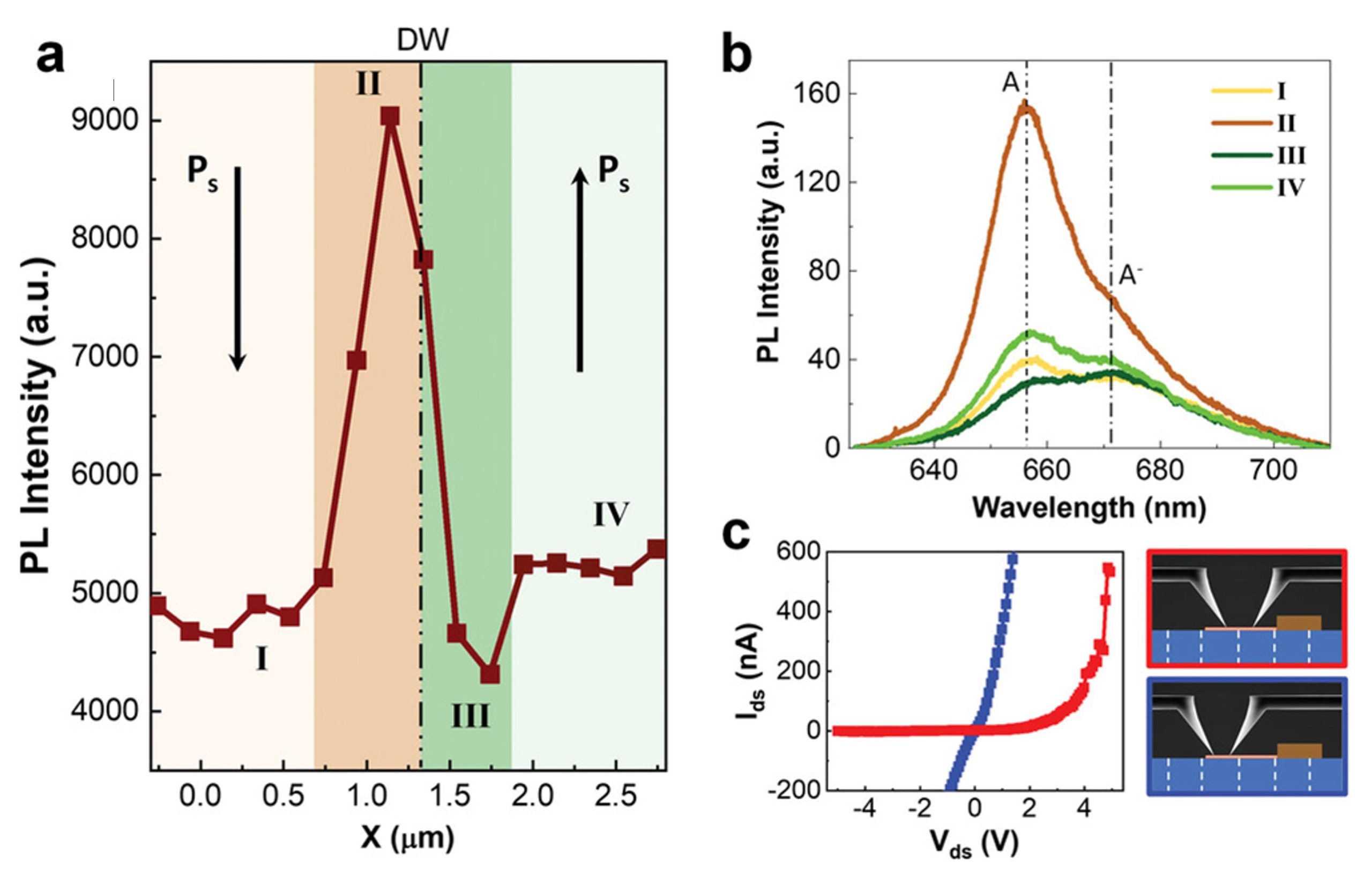
In the article “Light-Induced Ferroelectric Modulation of p-n Homojunctions in Monolayer MoS2” by Mariola O Ramirez, Jaime Fernandez-Tejedor, Daniel Gallego, Javier Fernández-Martinez, Pablo Molina, David Hernández-Pinilla, Julio Gómez-Herrero, Pablo Ares and Luisa E. Bausá, the combined effect of #ferroelectricity and light on the #optoelectronicproperties of monolayer (1L)-MoS2 deposited on periodically poled #lithiumniobate crystals is explored. *
Using scanning micro-photoluminescence, the effect of excitation intensity, scanning direction, and #domainwalls on the 1L-MoS2 #photoluminescence properties is analyzed, offering insights into charge modulation of #MoS2. *
The findings unveil a photoinduced charging process dependent on the #ferroelectricdomainorientation, in which light induces charge generation and transfer at the monolayer-substrate interface. *
This highlights the substantial role of light excitation in ferroelectrically-driven electrostatic doping in MoS2.
Additionally, the work provides insights into the effect of the strong, nanometrically confined electric fields on LiNbO3 domain wall surfaces, demonstrating precise control over charge carriers in MoS2, and enabling the creation of deterministic p-n homojunctions with exceptional precision.
The results suggest prospects for novel optoelectronic and photonic application involving monolayer TMDs by combining light-matter interaction processes and the surface selectivity provided by ferroelectric domain structures.
To corroborate the optical results, Mariola O Ramirez et al. measured current-voltage (I-V) curves by using two #AFMcantilevers with platinum-coated #AFMtips in contact with the 1L-MoS2 i) on a single domain region, and ii) on both sides of a ferroelectric domain wall where the p-n junction is formed. The results are shown in Figure 3c. (cited in here).
The #electricalcharacterization was carried out by means of a home-built two-terminal probe station with 2 sets of xyz piezomotors that allow precise positioning of the electrical probes.
NANOSENSORS™ AdvancedTEC™ ATEC-EFM #tipviewAFMprobes, conductive AFM tips that protrude from the very end of the AFM cantilever, ensuring real AFM tip visibility from above for a soft and accurate mechanical and electrical contact. *
To characterize the electrical properties of the system, the ATEC-EFM probes were brought into direct contact with the MoS2 flakes , as verified using a force sensor with a sensitivity of ≈1 mN located underneath the sample. This ensures a good electrical contact between the #AFMprobes and the MoS2. *
Full citation and direct link to the full article in the NANOSENSORS blog:

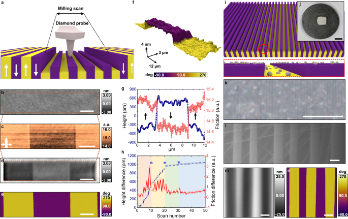
Switchable friction and wear behavior of ferroelectrics is observed with AFM.Mon Sep 02 2024
“These findings demonstrate that ferroelectrics are electrically tunable tribological materials at the nanoscale for versatile applications.”
Our gold coated 4XC-GG AFM probes and platinum coated HQ:DPER-XSC11 AFM probes are used for visualization and lithography of PbTiO3 thin films, PFM imaging and switching ferroelectric domains.
Morphological changes of plasma membrane and protein assembly during clathrin-mediated endocytosisFri Aug 30 2024
#Cells communicate with their environments via the #plasma #membrane and various #membrane #proteins. Clathrin-mediated endocytosis (CME) plays a central role in such communication and proceeds with a series of multiprotein assembly, deformation of the #plasmamembrane, and production of a membrane #vesicle that delivers extracellular signaling molecules into the cytoplasm.*
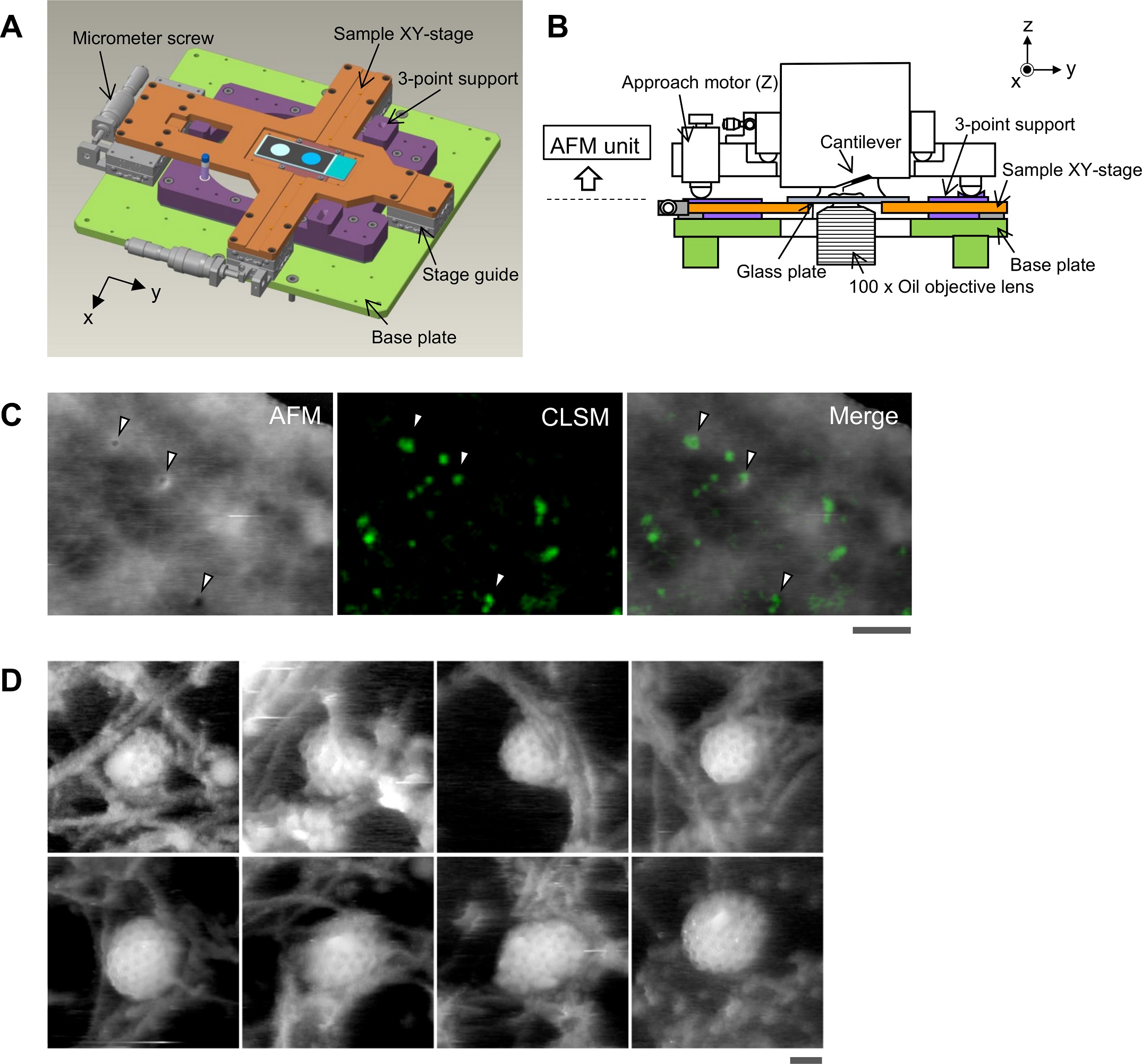
In the article “Morphological changes of plasma membrane and protein assembly during clathrin-mediated endocytosis”, Aiko Yoshida, Nobuaki Sakai, Yoshitsugu Uekusa, Yuka Imaoka, Yoshitsuna Itagaki, Yuki Suzuki and Shige H. Yoshimura describe how they utilized their home-built correlative imaging system comprising #highspeedatomicforcemicroscopy ( #HSAFM) and confocal #fluorescencemicroscopy to simultaneously image morphological changes of the plasma membrane and protein localization during CME in a #livingcell.*
Overlaying #AFM and fluorescence images revealed the dynamics of protein assembly and concomitant morphological changes of the plasma membrane with high spatial resolution. In particular, the authors elucidate the role of actin in the closing step of CME.*
The results revealed a tight correlation between the size of the pit and the amount of #clathrin assembled. #Actin dynamics play multiple roles in the assembly, maturation, and closing phases of the process, and affects membrane morphology, suggesting a close relationship between endocytosis and dynamic events at the cell cortex. Knock down of dynamin also affected the closing motion of the pit and showed functional correlation with actin.*
An AFM tip-scan–type HS-AFM unit combined with an inverted fluorescence/optical microscope equipped with a phase contrast system and a confocal unit was used for this study.*
The modulation method was set to phase modulation mode to detect AFM tip–sample interactions. A customized NanoWorld Ultra-Short #AFMcantilever with an electron beam–deposited sharp #AFMtip with a spring constant of 0.1 N m−1 (USC-F0.8-k0.1-T12) was used. *
Please have a look at the NanoWorld blog for the full citation and a direct link to the full article.


Structural Protein Changes Captured at High Temporal and Spatial ResolutionsFri Aug 30 2024
- Title: Deciphering the actin structure-dependent preferential cooperative binding of cofilin
- DOI: 10.1101/2023.12.06.570358
- Authors: Ngo, Kien Xuan and Vu, Huong T and Umeda, Kenichi and Trinh, Minh-Nhat and Kodera, Noriyuki and Uyeda, Taro Q.P.
- Publication: bioRxiv
- Pubisher: Cold Spring Harbor Laboratory
- Date: May 29, 2024

Stop by NanoAndMore USA booth 1407 at the ACSFALL2024Tue Aug 20 2024
Good Day Denver! #ACSFALL2024 has begun! What’s NEW in the #AFMProbe nanoverse? Stop by @NanoAndMore USA booth 1407 and see!
Expo Hours:
Monday & Tuesday
11AM - 5PM
Wednesday
10AM - 2PM
Exclusive hours for Posters and Expo Networking Break.
Quantifying mechanical forces during vertebrate morphogenesisThu Aug 15 2024
#Morphogenesis requires #embryoniccells to generate forces and perform mechanical work to shape their #tissues. Incorrect functioning of these #forcefields can lead to congenital malformations.*
Understanding these #dynamicprocesses requires the quantification and profiling of three-dimensional mechanics during evolving #vertebratemorphogenesis.*
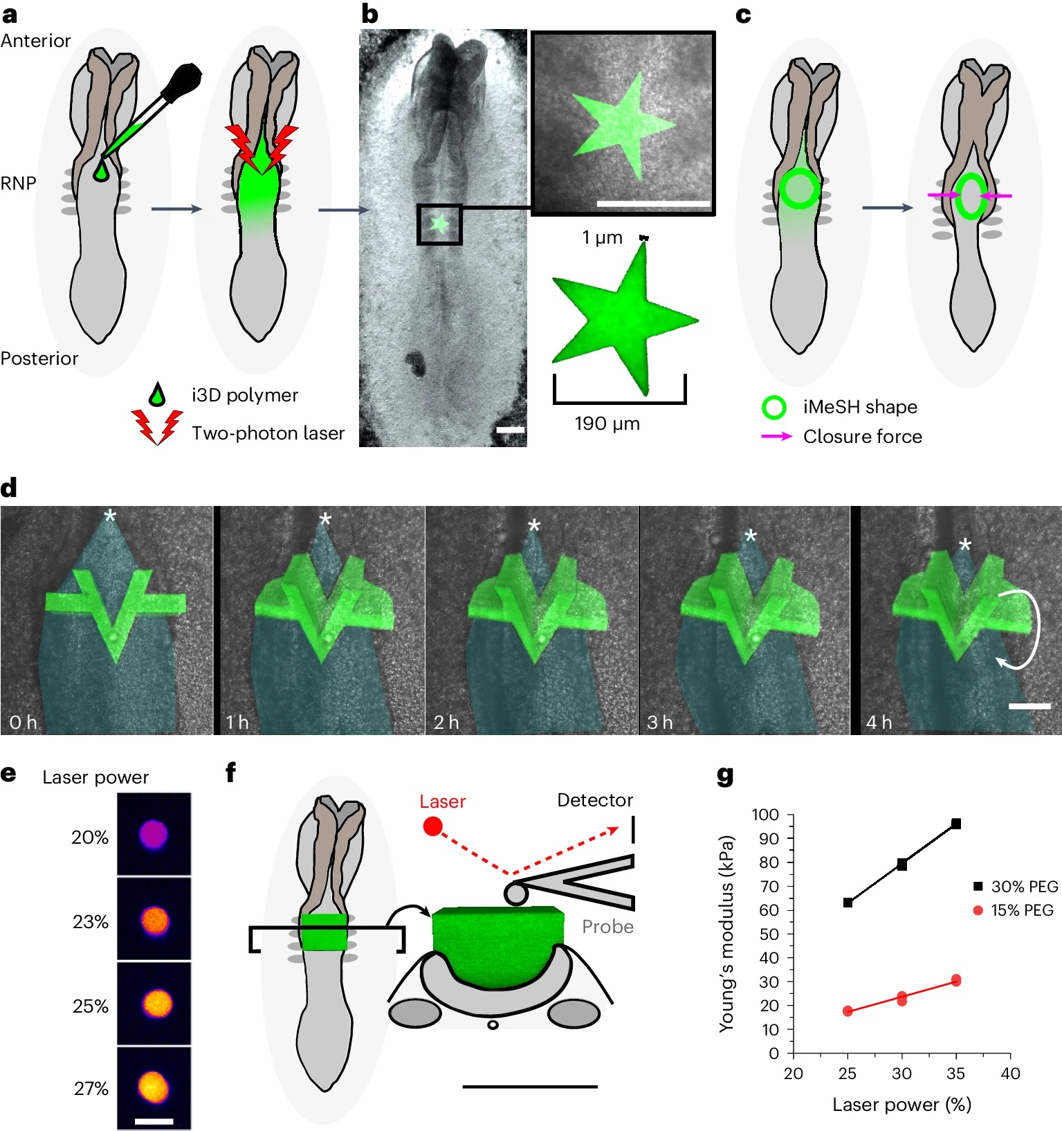
In the article “Quantifying mechanical forces during vertebrate morphogenesis” Eirini Maniou, Silvia Todros, Anna Urciuolo, Dale A. Moulding, Michael Magnussen, Ioakeim Ampartzidis, Luca Brandolino, Pietro Bellet, Monica Giomo, Piero G. Pavan, Gabriel L. Galea and Nicola Elvassore describe elastic spring-like #forcesensors with micrometre-level resolution, fabricated by intravital three-dimensional #bioprinting directly in the closing #neuraltubes of growing chicken #embryos.*
Integration of calibrated sensor read-outs with computational mechanical modelling allows direct quantification of the forces and work performed by the #embryonictissues. As they displace towards the embryonic midline, the two halves of the closing neural tube reach a compression of over a hundred nano-newtons during neural fold apposition. Pharmacological inhibition of Rho-associated kinase to decrease the pro-closure force shows the existence of active anti-closure forces, which progressively widen the neural tube and must be overcome to achieve neural tube closure. *
Overall, the author’s approach and findings highlight the intricate interplay between #mechanicalforces and #tissuemorphogenesis.*
The #atomicforcemicroscopy ( #AFM) described in the article was conducted using a commercially available #atomicforcemicroscope.*
The #forcedisplacementcurves were acquired using @NANOSENSORS @PointProbePlus PPP-CONTSCR silicon #AFMprobes with a typical spring constant of 0.2 N/m. *
The #AFMcantilever #springconstants were calibrated by the manufacturer prior to use. The sensitivity of each AFM cantilever was adjusted by measuring the slope of the #forcedistancecurve acquired on a hard reference material prior to each experiment. *
#Indentation experiments were repeated at least three times for each sample, at different locations. All AFM measurements were done in a fluid environment (PBS) at room temperature.*
The #Youngsmodulus was calculated by applying a fit of the Hertz model to the force–distance curve, assuming a Poisson ratio of 0.5, as is common practice for PEG #hydrogels. Preliminary in silico analyses of the #AFMtesting procedure were carried out to evaluate the effects of boundary conditions on the estimation of Young’s modulus.*
You will find the full citation and a direct link to the full article in the NANOSENSORS blog:

Piezoresponse Force Microscopy with BudgetSensors® ElectriMulti75-G AFM probesTue Aug 13 2024
Piezoresponse Force Microscopy with BudgetSensors® ElectriMulti75-G AFM probes supports this study comparing microwave assisted and conventional oven assisted hydrothermal syntheses of BaTiO3 nanoparticles for improved electroceramics.
Electrically and mechanically driven rotation of polar spirals in a relaxor ferroelectric polymerFri Aug 09 2024
Electrically and mechanically driven rotation of polar spirals in a relaxor ferroelectric polymer
Flux-closure structures, vortices/antivortices, skyrmions, and merons in oxides, metals and polymers represent non-trivial topologies in which a local polar/magnetic order undergoes quasi-continuous spatial variations in a host crystal lattice. These structures are now extensively studied due to emergent functionalities, but the application of electrical/mechanical fields has so far only served to destroy the polar topologies of interest. * Topology created by quasi-continuous spatial variations of a local polarization direction represents an exotic state of matter, but field-driven manipulation has been hitherto limited to creation and destruction. *In the article “Electrically and mechanically driven rotation of polar spirals in a relaxor […]
#AFMProbes, #ArrowCONTPt, #AtomicForceMicroscopy, #Characterization, #ConductiveAFMTip, #DomainWallCurvatures, #ElectronicMaterials, #ElectronicPropertiesAndMaterials, #FerroelectricPolymer, #FerroelectricPolymerThinFilms, #Ferroelectrics, #FerroelectricsAndMultiferroics, #InPlanePiezoResponseForceMicroscopy, #InPlanePiezoResponseForceMicroscopyIPPFM, #IPPFM, #MaterialsResearch, #MaterialsScience, #Multiferroics, #NéelRotation, #PFM, #PFMLithography, #PFMカンチレバー, #PFMプローブ, #PFM探针, #PiezoResponseForceMicroscopy, #PiezoresponseForceMicroscopy, #PolarSpiral, #PolarSpirals, #PolarizationMaps, #Polymers

Our booth for the #NCAFM2024 is all packed up and ready to travel home.Fri Aug 09 2024
Before we leave, we would like to say a big thank you to all of you who have visited our booth during the last few days.
And last but not least we would like to thank the local organizing committee at McGill University : Peter Grütter, Catherine Boisver, Louise Decelles, Chloé Paquet and Omur Dagdeviren (École de Technologie Supérieure) for a great conference.
We hope to see you all again soon!

Meet us at the 25th International Conference on Non-contact Atomic Force MicroscopyMon Aug 05 2024
Meet us @NanoAndMore #AFMprobes booth at the 25th International Conference on #NonContactAtomicForceMicroscopy from August 5th - August 9th at McGill University in Montreal this week. https://nc-afm2024.physics.mcgill.ca/
The conference covers the experimental, theoretical and instrumental developments in #frequencymodulation and other dynamic operation modes with particular emphasis on aspects of #highresolutionimaging and #forcespectroscopy.
We're hoping to see you soon!
Happy Birthday Switzerland – Swiss National Holiday 2024Wed Jul 31 2024
Happy Birthday Switzerland - Swiss National Holiday 2024
August 1st is the Swiss National Holiday. Enjoy the cheese fondue and have fun everyone!
Happy Birthday Switzerland!Wed Jul 31 2024
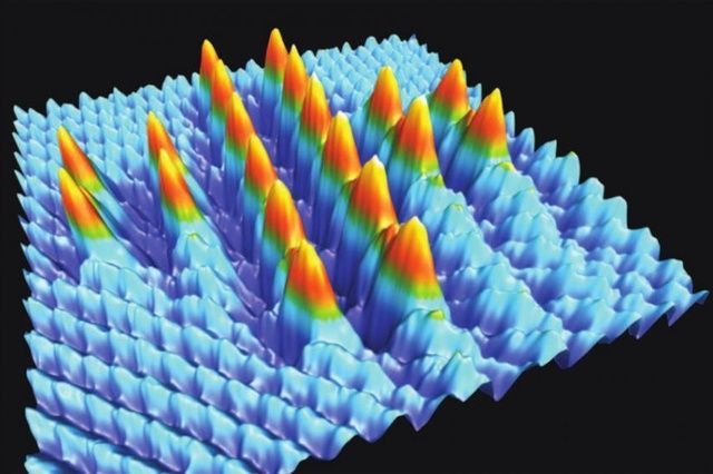
We are celebrating tomorrow’s #SwissNationalHoliday courtesy of Basel University ( @NanolinoBasel ) with the smallest #SwissCross – made of 20 single atoms.
Enjoy the holiday everyone in #Switzerland!
( a NANOSENSORS PointProbePlus PPP-NCL #AFMprobe was used for this image achieved with #atomicforcemicroscopy https://www.nanosensors.com/pointprobe-plus-non-contact… ).

HQ:NSC18/Pt AFM probes in Piezoresponce Force Microscopy (PFM) modeWed Jul 31 2024
Domain dynamics and resistive switching in ferroelectric Al(1-x)Sc(x)N thin film capacitors studied with our platinum coated HQ:NSC18/Pt AFM probes in Piezoresponce Force Microscopy (PFM) mode
The interface between ice and alcohols analyzed by atomic force microscopyMon Jul 29 2024
#Ice plays a crucial role in our environment, with natural ice formations, such as glaciers, permafrost, river ice, and snow, strongly influencing the temperature, humidity, and weather patterns on Earth.*
Because of its importance in our lives, extensive experimental and theoretical studies have been conducted to understand the characteristic properties of ice.*
An in-depth analysis of the interface between ice and water is essential for a complete understanding of ice near-natural conditions.*
In the article “The interface between ice and alcohols analyzed by atomic force microscopy” Ryo Yanagisawa, Tadashi Ueda, Keiichi Nakamoto , Zhengxi Lu, Hiroshi Onishi and Taketoshi Minato investigate the interface between ice and organic solvents using #atomicforcemicroscopy ( #AFM).*
Atomically flat ice surfaces were prepared and observed by AFM in 1-octanol, 1-hexanol, and 1-butanol.
The results show differences in #surfaceroughness influenced by the interaction of ice and alcohols.
The #Young’smodulus of ice was analyzed by #forcecurvemeasurements, providing valuable insights into the properties of ice in liquid environments.
The atomic force microscopy (AFM) measurements were conducted with a commercially available AFM that was placed in an acoustic enclosure and was cooled with the vapor of liquid nitrogen and a copper tube cooled with antifreezing fluid to maintain the environmental temperature at 264.7–270.2 K.
#Topographicimages were obtained in the #amplitudemodulationmode with NANOSENSORS PointProbe® Plus PPP-NCHAuD #AFMprobes with gold coating on the detector side of the #AFMcantilever.
The spring constant of each lever was calibrated from the Brownian motion of the AFM cantilever. To analyze Young’s modulus from the #forcecurve, the deflection sensitivity and #AFMtip radius were calibrated using Young’s modulus of mica (70 GPa).
To confirm the reproducibility of the atomic force microscopy results, the measurements were performed on 6–32 points (the distance between the points was more than 100 µm) from 3–16 ices under each condition.
Although the interface between alcohols and ice is different from that between water and ice, Ryo Yanagisawa et al. expect that careful selection of suitable organic solvents will lead to new insights that mimic essential features of the ice–water interface.*
Ryo Yanagisawa et al. they observed the surface structure of ice in liquid environments and demonstrated the analysis of physical properties, such as Young’s modulus, through force curve measurements in these liquid systems.
The results showed the characteristics of the ice surface in different solvents, suggesting potential applications in understanding surface and interface phenomena associated with ice under realistic conditions.*
Please have a look at the NANOSENSORS blog for the full citation and a direct link to the full article.

Improving Current and External Quantum Efficiency of Perovskite LEDsWed Jul 24 2024
Discover how nanotools conical SSS-NCHR with 2 nm radius are applied to study the topography of CsPbBr3 nanocrystals treated with hydrazine monohydrobromide.
- Title: Overcoming charge transfer barriers via electrostatically stabilized CsPbBr3 nanocrystals for efficient perovskite light-emitting diodes
- DOI: 10.1016/j.cej.2023.142120
- Authors: Min-Gi Jeon, Artavazd Kirakosyan, ChaeHo Shin, Subin Yun, Joonseok Kim, Li Li, Jihoon Choi
- Publication: Chemical Engineering Journal
- Publisher: Elsevier
- Date: 15 April 2023

Dielectric Properties of Polymer Nanocomposite Interphases Using Electrostatic Force Microscopy and Machine LearningMon Jul 22 2024
Metrology for measuring the interfacial dielectric permittivity in dielectrics with Electrostatic Force Microscopy (EFM) measurements and machine learning. AFM measurements performed using our platinum coated ElectriMulti75-G AFM probes.
Highlights


AFM Probe Focus


qp-HBC
uniqprobe™ - HeartBeat Cantilever for ScanAsyst®** and Peak Force Tapping™**
Coating:
Reflective Aluminum
Tip Shape: Circular symmetric
Tip Shape: Circular symmetric
AFM Cantilever:
F
60 kHz
C
0.5 N/m
L
115 µm
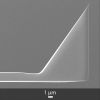
ATEC-NC
Tapping Mode AFM Probe with REAL Tip Visibility
Coating:
none
Tip Shape: Visible
Tip Shape: Visible
AFM Cantilever:
F
335 kHz
C
45 N/m
L
160 µm
TESPA
Standard Tapping Mode AFM Probe
Coating:
Reflective Aluminum
Tip Shape: Standard
Tip Shape: Standard
AFM Cantilever:
F
320 kHz
C
42 N/m
L
125 µm
OTESPA
Standard Tapping Mode AFM Probe with AFM Tip at the Very End of the AFM Cantilever
Coating:
Reflective Aluminum
Tip Shape: Optimized Positioning
Tip Shape: Optimized Positioning
AFM Cantilever:
F
300 kHz
C
26 N/m
L
160 µm