Soft, drift-reduced AFM cantilevers for Biology and Life Sciences – Uniqprobe Screencast passes the 1000 views markThu Aug 06 2020
The screencast on the soft, drift-reduced NANOSENSORS™ uniqprobe cantilevers for biology and life science applications held by Dr. Laure Aeschimann has just passed the 1000 views mark. Congratulations Laure!
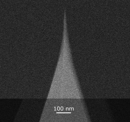
Since the first publication of this screencast that presents the uniqprobe types qp-BioAC, qp-BioT, qp-SCONT and qp-CONT , three further types of uniqprobe AFM probes have been introduced:
- qp-BioAC-CI – a version of uniqprobe™ BioAC with Rounded Tips for Cell Imaging
- qp-fast – three different uniqprobe™ cantilevers on one support chip for Soft- , Standard- , Fast Tapping/Dynamic AFM Imaging
and qp-HBC – the uniqprobe™ – HeartBeatCantilever
To find out more please have a look at the NANOSENSORS™ uniqprobe brochure https://www.nanosensors.com/…/3cfe8ca6ad48a762e2668b7d2b205… or the individual product pages on the NANOSENSORS webpage.
Additionally we have also put tipless versions of the qp-SCONT, qp-CONT and the qp-BioT ( SD-qp-BioT-TL, SD-qp-CONT-TL, SD-qp-SCONT-TL) and uniqprobe tipless cantilever arrays ( SD-qp-TL8a and SD-qp-TL8b ) on the NANOSENSORS special developments list. https://www.nanosensors.com/pdf/SpecialDevelopmentsList.pdf
Feel free to browse or let us know if you have any questions via info(at)nanosensors.com.


Influence of Ultraviolet Radiation Exposure Time on Styrene-Ethylene-Butadiene-Styrene (SEBS) CopolymerWed Aug 05 2020
In the article «Influence of Ultraviolet Radiation Exposure Time on Styrene-Ethylene-Butadiene-Styrene (SEBS) Copolymer» Daniel Garcia-Garcia , José Enrique Crespo-Amorós, Francisco Parres and María Dolores Samper describe how they studied the effect of ultraviolet radiation on styrene-ethylene-butadiene-styrene (SEBS) at different exposure times in order to obtain a better understanding of the mechanism of ageing.*
The results obtained for the SEBS, in relation to the duration of exposure, showed superficial changes that cause a decrease in the surface energy (s) and, therefore, a decrease in surface roughness. This led to a reduction in mechanical performance, decreasing the tensile strength by about 50% for exposure times of around 200 hours.*
NanoWorld Pointprobe® NCH AFM probes were used in the Atomic force microscopy (AFM) applied to determine the surface topography and roughness of the aged samples. https://www.nanoworld.com/pointprobe-tapping-mode-afm-tip…
Please have a look at the NanoWorld blog for the full citation and a direct link to the full article.

Direct Measurement of Adhesion Force of Individual Aerosol Particles by Atomic Force MicroscopyTue Aug 04 2020
In the article «Direct Measurement of Adhesion Force of Individual Aerosol Particles by Atomic Force Microscopy» Kohei Ono, Yuki Mizushima, Masaki Furuya, Ryota Kunihisa, Nozomu Tsuchiya, Takeshi Fukuma, Ayumi Iwata and Atsushi Matsuki describe a new method, namely, force–distance curve mapping, that was developed to directly measure the adhesion force of individual aerosol particles by atomic force microscopy.*
The proposed method collects adhesion force from multiple points on a single particle. It also takes into account the spatial distribution of the adhesion force affected by topography (e.g., the variation in the tip angle relative to the surface, as well as the force imposed upon contact), thereby enabling the direct and quantitative measurement of the adhesion force representing each particle.*
The results presented in the article highlight that the original chemical composition, as well as the aging process in the atmosphere, can create significant variation in the adhesion force among individual particles. This study demonstrates that force–distance curve mapping can be used as a new tool to quantitatively characterize the physical properties of aerosol particles on an individual basis.*
The measurement of adhesion force described in the article was performed in contact mode using silicon NANOSENSORS™ AdvancedTEC™ ATEC-CONT AFM tips.*
Please have a look at the NANOSENSORS blog for the full citation and a direct link to the full article.


Determination of polarization states in (K,Na)NbO3 lead-free piezoelectric crystalTue Aug 04 2020
Check out this article describing how polarization switching in lead-free (K0.40Na0.60)NbO3 (KNN) single crystals was studied by switching spectroscopy piezoresponse force microscopy (SS-PFM).
PFM and SS-PFM were implemented on a commercial Atomic Force Microscope using NanoWorld PlatinumIridium coated Pointprobe® EFM AFM probes. https://www.nanoworld.com/pointprobe-electrostatic-force…


MikroMasch rotated AFM tipsMon Jul 27 2020
When mounted at the standard 13⁰ angle in the AFM, the front and back edges of MikroMasch rotated AFM tips form the same 20⁰ angle to the vertical. This allows more symmetric visualization of topography features than when using non-rotated tips.


Happy birthday to Gerd BinnigMon Jul 20 2020
Happy birthday to Nobel prize laureate Gerd Binnig, coinventor of the Scanning Tunneling Microscope
and the Atomic Force Microscope!

Nanotools biosphere™ AFM probes applied to study the mechanical properties of #PDMS filmsWed Jul 15 2020
Discover how nanotools biosphere™ AFM probes with precisely controlled 500 nm radius and pre-calibrated cantilevers is applied to study the mechanical properties of #PDMS films in the latest nanotools blogpost


Gentle plasma process for embedded silver-nanowire flexible transparent electrodes on temperature-sensitive polymer substratesTue Jul 14 2020
In the article “Gentle plasma process for embedded silver-nanowire flexible transparent electrodes on temperature-sensitive polymer substrates “ Lukas Kinner, Emil J W List-Kratochvil and Theodoros Dimopoulos investigate processing routes to obtain highly conductive and transparent electrodes of silver nanowires (AgNWs) on flexible polyethylene terephthalate (PET) substrate.*
Their study shows that both thermally stable polyimide, as well as temperature-sensitive PET can be used as flexible host substrates, combined with a gentle, AgNW plasma curing. This is possible by adjusting the fabrication sequence to accommodate the plasma curing step, depending on the host substrate. As a result, embedded AgNW electrodes, transferred from polyimide-to-PET and from PET-to-PET are obtained, with optical transmittance of ~80% (including the substrate) and sheet resistance of ~13 Ω/sq., similar to electrodes transferred from glass-to-glass substrates.*
The embedded AgNW electrodes on PET show superior performance in bending tests, as compared to indium-tin-oxide electrodes and can be easily combined with metal oxide films for device implementation. The introduced approach, involving low-cost flexible substrates, AgNW spray-coating and plasma curing, is compatible with high-throughput, roll-to-roll processing.*
The impact of the introduced processes concerns therefore applications where high-throughput production must be combined with sensitive, flexible substrates and ultra-thin device architectures, like OLEDs and organic- or perovskite-based photovoltaics.*
The sample surfaces were characterized with atomic force microscopy (AFM) in tapping mode, using high-resolution NANOSENSORS™ SuperSharpSilicon™ SSS-NCHR AFM probes. https://www.nanosensors.com/supersharpsilicon-non-contact…
Please have a look at the NANOSENSORS blog for the full citation and a direct link to the full article


Human ESCRT-III polymers assemble on positively curved membranes and induce helical membrane tube formationMon Jul 06 2020
The Endosomal Sorting Complex Required for Transport-III (ESCRT-III) is part of a conserved membrane remodeling machine. ESCRT-III employs polymer formation to catalyze inside-out membrane fission processes in a large variety of cellular processes, including budding of endosomal vesicles and enveloped viruses, cytokinesis, nuclear envelope reformation, plasma membrane repair, exosome formation, neuron pruning, dendritic spine maintenance, and preperoxisomal vesicle biogenesis.*
How membrane shape influences ESCRT-III polymerization and how ESCRT-III shapes membranes is yet unclear.*
In the article “Human ESCRT-III polymers assemble on positively curved membranes and induce helical membrane tube formation” Aurélie Bertin, Nicola de Franceschi, Eugenio de la Mora, Sourav Maity, Maryam Alqabandi, Nolwen Miguet, Aurélie di Cicco, Wouter H. Roos, Stéphanie Mangenot, Winfried Weissenhorn and Patricia Bassereau describe how human core ESCRT-III proteins, CHMP4B, CHMP2A, CHMP2B and CHMP3 are used to address this issue in vitro by combining membrane nanotube pulling experiments, cryo-electron tomography and Atomic Force Microscopy.*
The authors show that CHMP4B filaments preferentially bind to flat membranes or to tubes with positive mean curvature.*
The results presented in the article cited above underline the versatile membrane remodeling activity of ESCRT-III that may be a general feature required for cellular membrane remodeling processes.*
The authors provide novel insight on how mechanics and geometry of the membrane and of ESCRT-III assemblies can generate forces to shape a membrane neck.*
NanoWorld Ultra-Short AFM Cantilevers USC-F1.2-k0.15 were used for the High-speed Atomic Force Microscopy ( HS-AFM ) experiments presented in this article.* https://www.nanoworld.com/Ultra-Short-Cantilevers-USC-F1.2…
Please have a look at the NanoWorld blog for the full citation and a direct link to the full article.


A two-step method for rate-dependent nano-indentation of hydrogelsFri Jul 03 2020
Two-step method for accurate alignment of finite-rate nanoindentation curves on hydrogels using a colloidal AFM probe based on our MikroMasch NSC36/tipless/Cr-Au

Studying nanomechanical properties of primary cell walls in the inner tissues of growing plant organsMon Jun 29 2020
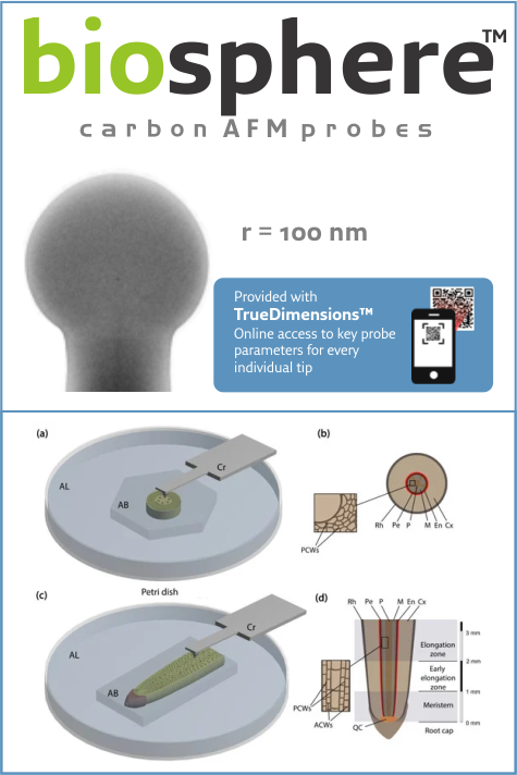
Discover how nanotools biosphere™ #AFMprobes with precisely controlled 100 nm radius and pre-calibrated cantilevers are applied for imaging and nanoindentation of plant cell walls in the latest nanotools blogpost https://www.nanotools.com/…/studying-nanomechanical…


Development of a Lidocaine-Loaded Alginate/CMC/PEO Electrospun Nanofiber Film and Application as an Anti-Adhesion BarrierThu Jun 25 2020
Surgery, particularly open surgery, is known to cause tissue/organ adhesion during healing. These adhesions occur through contact between the surgical treatment site and other organ, bone, or abdominal sites. Fibrous bands can form in unnecessary contact areas and cause various complications. Consequently, film- and gel-type anti-adhesion agents have been developed. The development of sustained drug delivery systems is very important for disease treatment and prevention.*
In “Development of a Lidocaine-Loaded Alginate/CMC/PEO Electrospun Nanofiber Film and Application as an Anti-Adhesion Barrier” Seungho Baek, Heekyung Park, Youngah Park, Hyun Kang and Donghyun Lee describe how the drug release behavior was controlled by crosslinking lidocaine-loaded alginate/carboxymethyl cellulose (CMC)/polyethylene oxide (PEO) nanofiber films prepared by electrospinning.*
Lidocaine is mainly used as an anesthetic and is known to have anti-adhesion effects.*
Based on the results presented in the article, this study shows that the drug release behavior can be controlled by using CaCl2 as a nontoxic crosslinking agent to produce a good anti-adhesion barrier that can prevent unnecessary tissue adhesion at a surgical site.*
The authors selected atomic force microscopy (AFM) using NANOSENSORS™ PointProbe® Plus PPP-NCHR AFM cantilevers to analyze the electrospun films.* https://www.nanosensors.com/pointprobe-plus-non-contact…
Please have a look at the NANOSENSORS blog for the full citation and a direct link to the full article.

BudgetSensors® SiNi AFM probes used to examine cell stiffness.Wed Jun 24 2020
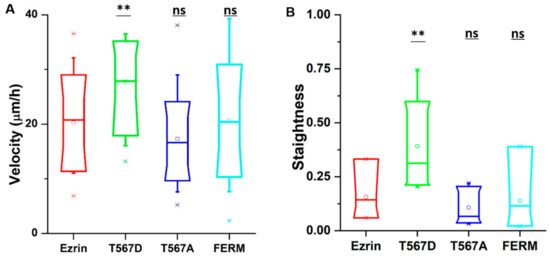
Тhe role of ezrin phosphorylation on cell motility, cytoskeleton organization and mechanical properties. BudgetSensors® SiNi AFM probes used to examine cell stiffness.


Chemical switching of low-loss phonon polaritons in α-MoO3 by hydrogen intercalationTue Jun 23 2020
Chemical switching of low-loss phonon polaritons in α-MoO3 by hydrogen intercalation
Phonon polaritons (PhPs) have attracted significant interest in the nano-optics communities because of their nanoscale confinement and long lifetimes. Although PhP modification by changing the local dielectric environment has been reported, controlled manipulation of PhPs by direct modification of the polaritonic material itself has remained elusive.*
In the article “Chemical switching of low-loss phonon polaritons in α-MoO3 by hydrogen intercalation” Yingjie Wu, Qingdong Ou, Yuefeng Yin, Yun Li, Weiliang Ma, Wenzhi Yu, Guanyu Liu, Xiaoqiang Cui, Xiaozhi Bao, Jiahua Duan, Gonzalo Álvarez-Pérez, Zhigao Dai, Babar Shabbir, Nikhil Medhekar, Xiangping Li, Chang-Ming Li, Pablo Alonso-González and Qiaoliang Bao demonstrate an effective chemical approach to manipulate PhPs in α-MoO3 by the hydrogen intercalation-induced perturbation of lattice vibrations.*
Their methodology establishes a proof of concept for chemically manipulating polaritons, offering opportunities for the growing nanophotonics community.*
The surface topography and near-field images presented in this article were captured using a commercial s-SNOM setup with a platinum iridium coated NanoWorld Arrow-NCPt AFM probe in tapping mode.* https://www.nanoworld.com/tapping-mode-platinum-coated-afm…
Please have a look at the Nanoworld blog for the full citation and a direct link to the full article


Combined SIM and AFM platform for the life sciencesMon Jun 22 2020
Correlating data from different microscopy techniques holds the potential to discover new facets of signalling events in cellular biology.*
In the article “Simultaneous co-localized super-resolution fluorescence microscopy and atomic force microscopy: combined SIM and AFM platform for the life sciences” Ana I. Gómez-Varela, Dimitar R. Stamov, Adelaide Miranda, Rosana Alves, Cláudia Barata-Antunes, Daphné Dambournet, David G. Drubin, Sandra Paiva and Pieter A. A. De Beule report for the first time a hardware set-up capable of achieving simultaneous co-localized imaging of spatially correlated far-field super-resolution fluorescence microscopy and atomic force microscopy, a feat only obtained until now by fluorescence microscopy set-ups with spatial resolution restricted by the Abbe diffraction limit.*
The authors detail system integration and demonstrate system performance using sub-resolution fluorescent beads and applied to a test sample consisting of human bone osteosarcoma epithelial cells, with plasma membrane transporter 1 (MCT1) tagged with an enhanced green fluorescent protein (EGFP) at the N-terminal.*
The simultaneous operation of AFM and super-resolution fluorescence microscopy technique provides a powerful observational tool on the nanoscale, albeit data acquisition is typically obstructed by a series of integration problems. The authors of the above-mentioned article believe that the combination of SR-SIM with AFM presents one of the most promising schemes enabling simultaneous co-localized imaging, allowing the recording of nanomechanical data and cellular dynamics visualization at the same time.*
For measurements on cells in liquid NANOSENSORS™ uniqprobe qp-BioAC-CI AFM probes ( CB1 ) with a nominal resonance frequency of 90 kHz (in air), spring constant of 0.3 Nm−1, partial gold coating on the detector side, and quartz-like circular symmetric hyperbolic (double-concaved) tips with ROC of 30 nm were used. The corresponding AFM areas for the cell images were acquired with a Z-cantilever velocity of 250 μms−1 at a max Z-length of 1.5 μm, resulting in an acquisition time (based on the number of pixels) for Figs. 2, 3, 4 of ca. 13, 8 and 15 min respectively.* https://www.nanosensors.com/uniqprobe-uniform-quality-bioac…
Please have a look at the NANOSENSORS blog for the full citation and a direct link to the full article

How to search for AFM probes?Fri Jun 19 2020
Check out our latest video
Nick Schacher is explaining the basic factors to consider before buying AFM probes.
Where do I find a TRUSTED Brand?
Where do I find the BEST Probe for my Application?
Where do I find a Probe that meets my Budget?
Why NanoAndMore?
MOST Trusted ~ 30+ Years of Business and Peer-Reviewed Papers!
MOST Options ~ 10 Different Brands in Stock – Same day shipment!
BEST Prices ~ Brands structured for ALL Budgets!
TOP Experience ~ 30+ Years of Expertise! Questions? Contact us!
FREE Probe Samples ~ TRY before you BUY!


NanoAndMore goes South East AsiaWed Jun 17 2020


Probing Mid-Infrared Phonon PolaritonsTue Jun 09 2020
Probing Mid-Infrared Phonon Polaritons in the Aqueous Phase with MikroMasch HQ:NSC14/Cr-Au


Nanoparticle characterizationThu May 28 2020
Nanoparticle characterization using TERS and Force-Distance based AFM with the help of the tried-and-true BudgetSensors Tap190Al-G and Tap150Al-G AFM probes


